Medical 3D-Printed Surgical Guides: From Patient Imaging to Manufacturable CAD
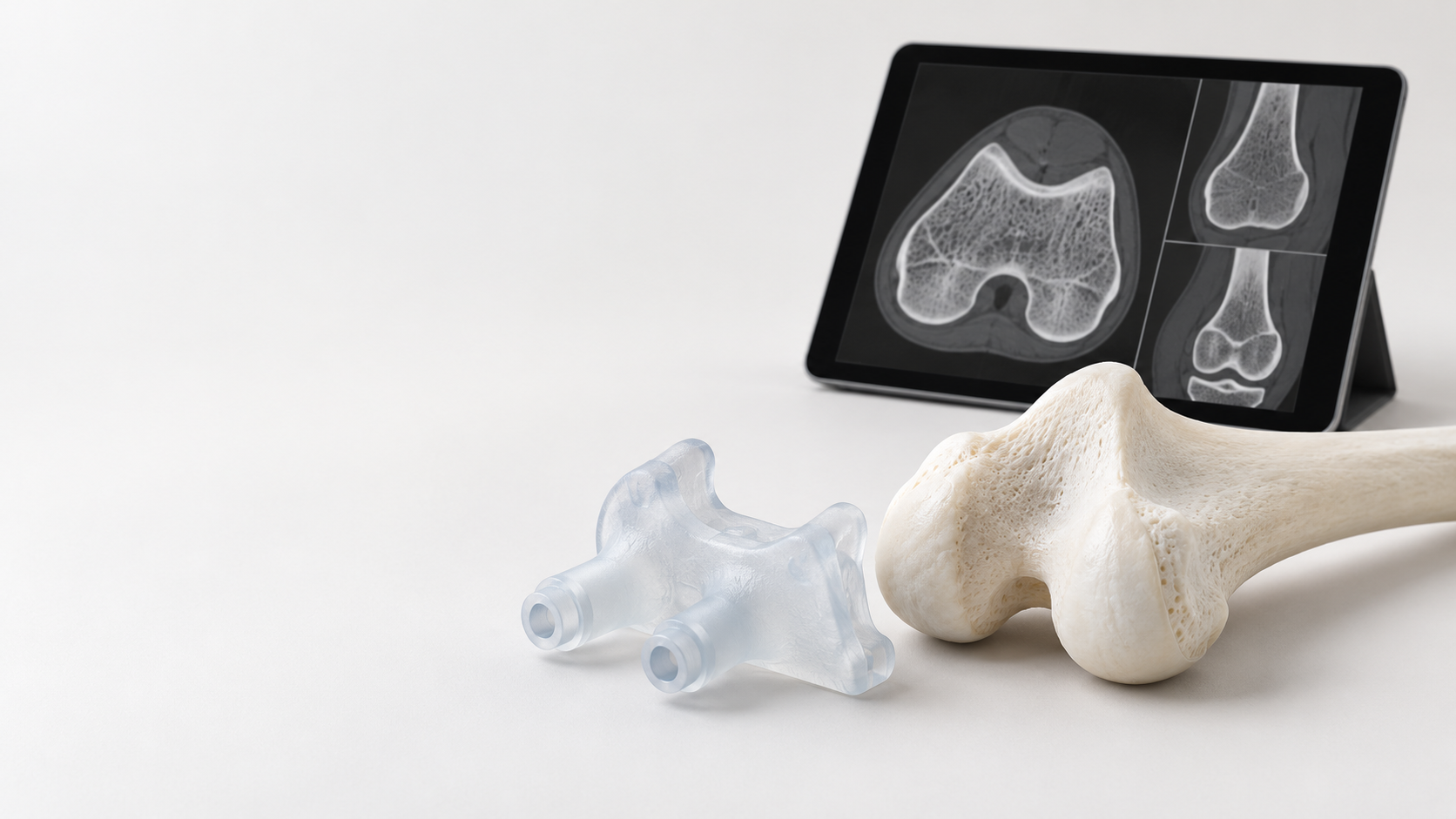
A surgical guide is not simply an ordinary prototype made with “medical-grade material.” It typically begins with the patient’s CT or other imaging data, undergoes data segmentation, 3D reconstruction, CAD design, and 3D printing, and must ultimately demonstrate that the finished product can position or guide instruments as intended. Therefore, the manufacturing accuracy of a medical 3D-printed surgical guide depends not only on the printing equipment but also on image quality, segmentation boundaries, design datums, material deformation, and post-processing.
How Medical 3D-Printed Surgical Guides Move from DICOM Data to 3D Models
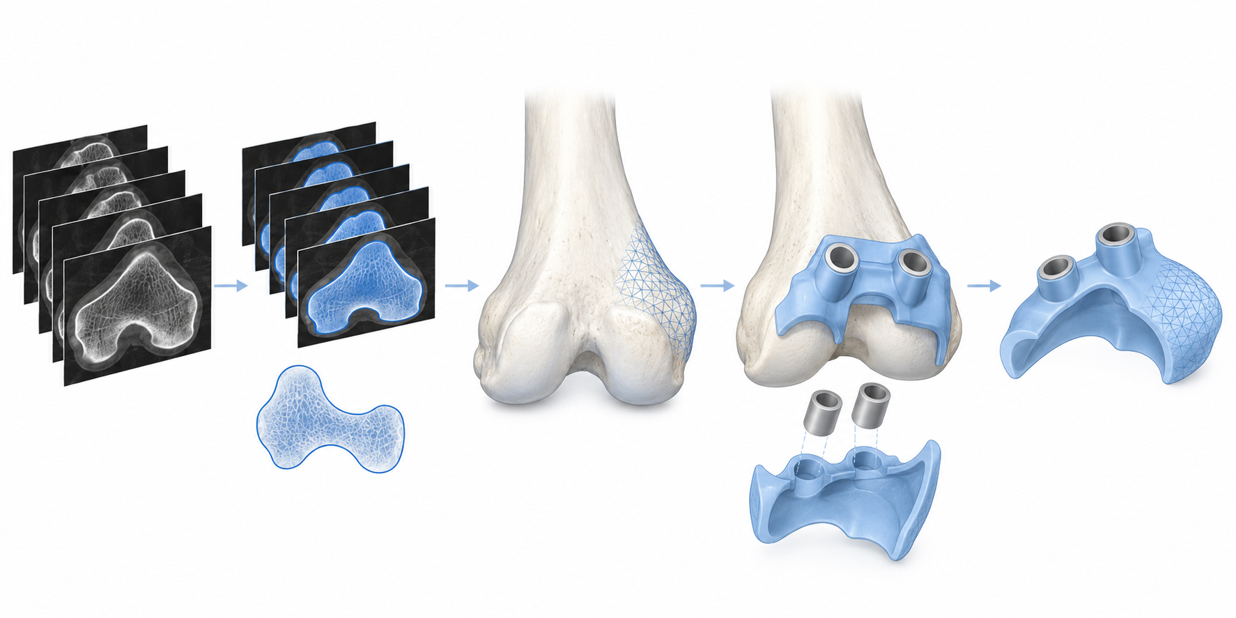
The first step is to confirm the original images, patient data, and clinical objectives, and then segment the DICOM data into the target bone surface or relevant anatomical structures. Segmentation thresholds, inter-slice spacing, artifacts, and boundary interpretation can all alter the 3D model; consistency between the model and the images does not necessarily mean that the subsequently printed part will be correct. The original images, segmentation files, 3D reconstruction models, and version information should be retained upon delivery to prevent outdated data from being used inadvertently during design.
Contact Surfaces and Guiding Features in Medical 3D-Printed Surgical Guide Design
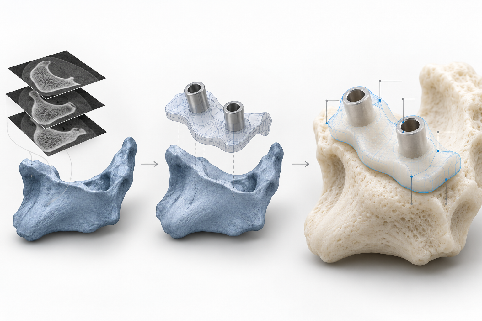
Using a bone-fitted drilling guide as an example, the CAD design must first define the bone-contact areas, followed by the placement of guide holes, locating steps, openings, and necessary viewing spaces. The contact surfaces should be positioned as much as possible using stable anatomical features as the locating datum; the guide holes must simultaneously account for hole diameter, axis, instrument clearance, and surrounding wall thickness. Thin walls, deep holes, and narrow openings may increase the difficulty of printing, cleaning, or inspection, so the form cannot be verified solely on screen.
After the design is completed, the versions, units, scale, and critical features of the CAD model and the STL or other manufacturing files used for printing should be checked to ensure that the model has not been flipped, scaled, or locally modified. Parameters such as device pixel size and layer thickness can serve only as references for process development and cannot be directly treated as the final tolerance of the guide. At this stage, traceable model-comparison and approval records should be created so that subsequent printing and inspection remain linked to the original patient data through a consistent version chain.
How to Select Materials for Medical 3D-Printed Surgical Guides: What Else to Consider Beyond Biocompatibility
The material selection for surgical guides should not begin with “which resin is the hardest” or “which material is labeled for medical use.” Instead, the intended use must first be defined: what tissue the guide will contact, how long it will be used, what positioning or drilling loads it will withstand, and which cleaning and sterilization methods will ultimately be used. For single-use, short-duration, bone-surface-fitting guides, biocompatibility, stiffness, dimensional stability, transparency, and surface residues generally all require attention. If the guide needs to undergo high-temperature, high-pressure sterilization, it must also be confirmed that the material will not soften significantly, warp, or lose positioning accuracy under the specified temperature, pressure, duration, and number of cycles.
Medical 3D printing materials should be compared by application scenario
Photocurable medical resins are suitable for reproducing small guide holes, thin walls, and complex conformal surfaces, but their performance is affected by cleaning, post-curing, and residual unreacted material. Transparency may also change after subsequent processing and sterilization. Powder-bed polymer processes such as nylon generally require focused evaluation of surface porosity, cleaning difficulty, dimensional stability, and guide-hole edge quality. Biocompatibility, mechanical stability, or sterilization statements provided by a material supplier for a specific grade can serve only as a basis for material selection; they do not automatically prove that the printed part has completed cleaning, sterilization, packaging, and clinical release validation.
- [object Object]
- [object Object]
- [object Object]
- [object Object]
Therefore, the material-selection conclusion for a medical 3D-printed surgical guide should be written as “a specific material grade, printing process, post-processing method, and sterilization condition are suitable for the intended use,” rather than simply stating that “the resin can be sterilized” or that “a biocompatible material can be used directly.” If the sterilization method has not yet been determined, the validation conditions should first be fixed during the quotation and design stages, followed by determination of the wall thickness, guide-hole structure, and surface requirements.
Where does the dimensional accuracy of a 3D-printed surgical guide come from?
The accuracy of a 3D-printed surgical guide is not a single number listed in a printer specification sheet. It involves at least three relationships: whether the original images are consistent with the anatomical model, whether the printed part conforms to the CAD model, and whether the guide can be positioned stably on the target bone surface. A deviation at any one of these levels may change the final position of the guide holes.
Equipment resolution is not the same as finished-part tolerance
For photopolymerization equipment, pixel size or spot size, layer thickness, and build volume affect the ability to reproduce details; however, print orientation, placement, support layout, exposure parameters, and material shrinkage are equally important. Guide holes, thin walls, openings, and bone-contact areas are particularly sensitive to these variations. The resolution stated in equipment or supplier documentation can serve only as a development reference and cannot be directly specified as the acceptance tolerance for a surgical guide. Engineering projects sometimes use equipment or process specifications at the scale of tens to hundreds of micrometers as an initial reference, but the final values must still be confirmed based on the specific material, geometric structure, and measurement system.
Three types of consistency must be checked separately for a 3D-printed surgical guide
| Inspection level | Main issues | Recommended evidence |
|---|---|---|
| Imaging to model | Whether resolution, segmentation, and anatomical boundaries cause deviations | Original images, segmentation model, and version records |
| CAD to finished part | Whether printing, shrinkage, support structures, and post-curing alter critical dimensions | Model comparison and critical-dimension measurement |
| From the finished part to the bone surface | Whether the contact surface is stable and whether the guide-hole axis meets the design intent | Fit, positioning, and functional inspection |
Therefore, a guide cannot be judged acceptable based on a single measurement of its outer profile. Priority should be given to verifying the bone-contact surface, guide-hole diameter and axis, locating steps, and thin-wall regions. For bone-contact drill guides, dimensional inspection confirms the relationship between the part and the digital model; fit inspection confirms the relationship between the part and the target anatomical surface. The two cannot substitute for each other.
Why post-processing of 3D-printed surgical guides must be included in verification
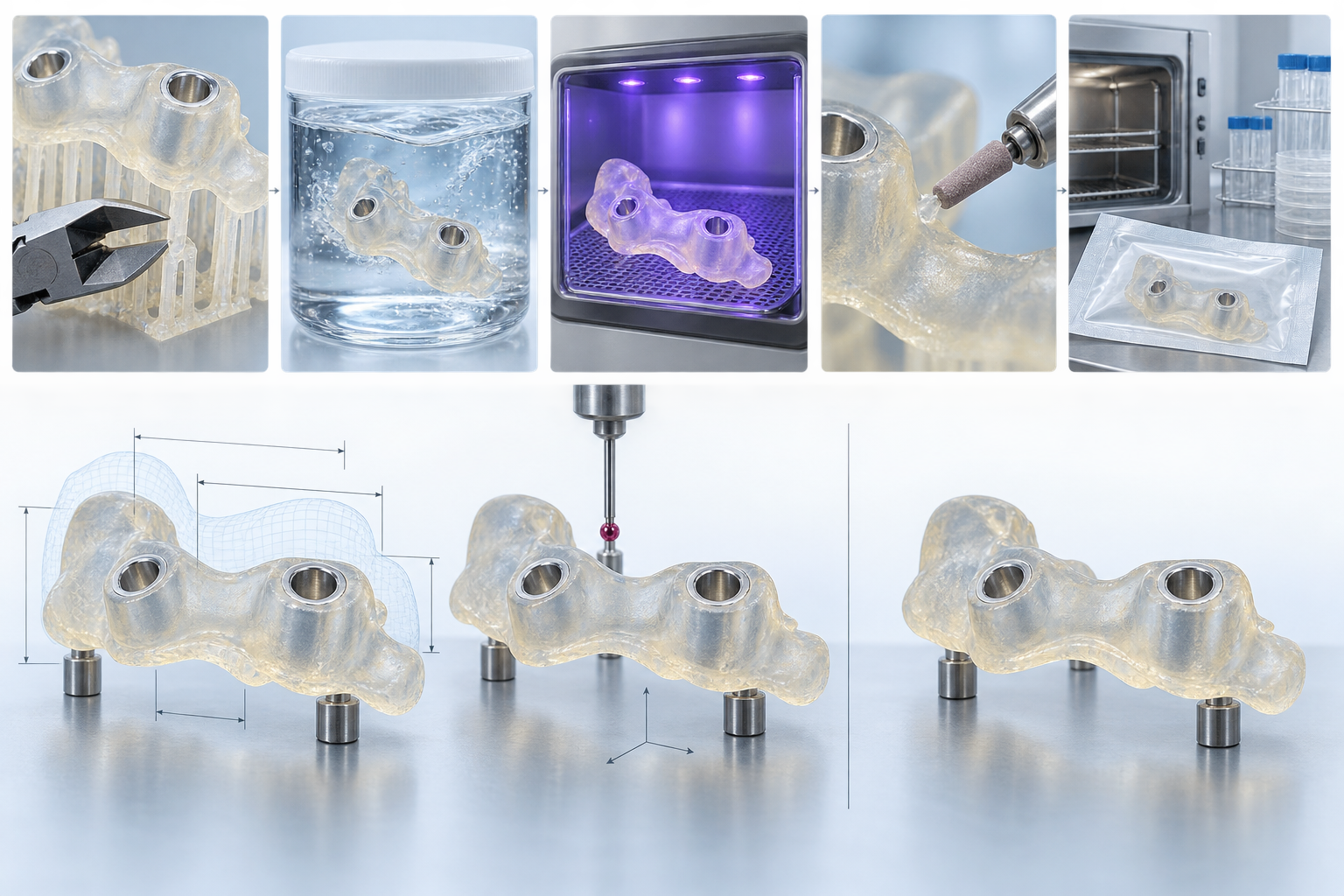
A surgical guide is not a finished part as soon as it leaves the printer. Support removal, cleaning, post-curing, surface treatment, and sterilization can all change the guide's dimensions, surface condition, or mechanical properties. For bone-contact drill guides, the locating surfaces, guide holes, and thin-wall regions are functional features, and any local deformation during post-processing may affect positioning stability. Therefore, post-processing in medical 3D printing should be treated as a manufacturing operation rather than a simple cleaning step.
Support removal and cleaning should first control local interference and residue
When removing supports, priority should be given to protecting the bone-contact areas, guide-hole edges, and locating steps. Protrusions left by support points may cause local interference, while excessive trimming may alter the fit surface or hole-mouth geometry. For photopolymerization processes, attention should also be paid to uncured material in internal holes, recesses, and narrow gaps; insufficient cleaning can result in residue, a tacky surface, or uneven subsequent curing. The cleaning medium, duration, flow method, and drying conditions should be confirmed according to the material and equipment process, with batch records retained.
Post-curing and surface treatment may alter critical dimensions
Post-curing continues to affect material shrinkage, stiffness, and brittleness, with time, energy, part orientation, and wall thickness all influencing the result. Sanding or polishing should not be evaluated by appearance alone: thinning the bone-contact area, enlarging guide holes, or rounding thin-wall edges may alter function. Reproducible parameter windows should be established for critical materials and geometries and confirmed using worst-case specimens containing thin walls, holes, recesses, and fit surfaces, rather than by validating only regular test coupons.
Sterilization validation must compare the condition before and after processing
If the guide requires sterilization, the material must be evaluated under the specified temperature, pressure, duration, and number of cycles for warpage, softening, cracking, or changes in positioning accuracy. A material supplier's sterilization statement for a specific product can serve only as a material-selection basis and cannot replace finished-product validation. In engineering practice, the bone-contact surface, guide-hole diameter and axis, locating steps, and external condition should be compared before and after sterilization; fit or instrument-guidance checks should be added when necessary. Final records should link the material lot number, printing parameters, post-processing parameters, sterilization batch, and inspection results so that any anomaly can be traced to a specific operation.
How to perform quality verification and release of personalized medical surgical guides
Quality verification of a surgical guide cannot be based solely on its appearance after printing or on a few dimensional measurements. For bone-contact drill guides, release criteria should cover six aspects: documentation, process, part, function, cleaning, and sterilization. Dimensional conformity does not mean that the guide can be positioned stably on the target bone surface, and a material supplier's biocompatibility statement does not mean that the finished product has completed cleaning, sterilization, and intended-use validation.
Verify Files and Processes First for Medical 3D-Printed Surgical Guides
At the file level, verify the original imaging data, segmentation model, patient or project number, CAD version, and whether the exported STL or print file corresponds to the same version. Any modification to a bone-surface contact area, guide-hole axis, or locating step should be documented with a version record and approval basis. At the process level, record the material grade and lot number, equipment, print orientation, layer thickness, placement, support strategy, cleaning and post-curing parameters. This is the only way to trace deviations in the finished product back to the model, material, or process stage, rather than simply repeating the print.
Critical Dimensions of Surgical Guides Must Also Undergo Functional Inspection
Part inspection should focus on functional features: measure the bone-surface contact areas, guide-hole diameter and axis, locating steps, thin walls, openings, and instrument clearance. Depending on the geometry, inspection methods may include gauges, 3D scanning, or comparison with a digital model, but the measurement system’s resolution, datum establishment method, and uncertainty must match the acceptance requirements. For complex surfaces, one overall dimension cannot be used as a substitute for checking the complete position and orientation.
Functional inspection should confirm whether the guide conforms to the design datums, whether positioning is stable, whether the guide holes allow the specified instruments to pass, and whether interference is introduced after support removal or surface treatment. Where necessary, compare the critical dimensions, warpage, and fit before and after sterilization. The final release record should also include the cleaning status, sterilization method and batch, packaging identification, model version, inspection results, and the name of the person making the disposition.
- File: The original imaging, segmentation model, CAD, and print-file versions are consistent.
- Process: The material lot number, equipment parameters, print orientation, and post-processing records are complete.
- Part: Critical dimensions, axes, thin walls, and surface condition meet product-specific requirements.
- Function: Bone-surface fit, positioning, and instrument guidance checks are complete.
- Release: Cleaning, sterilization, packaging, and traceability information can be linked to the individual product.
Therefore, a personalized medical surgical guide should not use a single uniform “±X mm” as the acceptance criterion for everything. A more robust approach is to establish product-specific acceptance criteria for this type of guide based on clinical risk, critical features, the material and process window, and actual measurement capabilities, while retaining verifiable release evidence.
How to Communicate with a 3D Printing Supplier for a Surgical Guide Manufacturing Project
A surgical guide is not an ordinary prototype that can be completed simply by uploading an STL file. The 3D printing supplier needs to know its intended use, contacted tissue, duration of use, and sterilization method. These factors directly affect the material, print orientation, post-processing, and final acceptance. Bone-fitting drill guides should also specify the guide-hole axes, locating surfaces, openings, thin-wall areas, and instrument entry direction.
Define the Intended Use and Acceptance Boundaries in the RFQ Documents First
Procurement documents should include at least the version information for the imaging data or model, the final CAD version, material restrictions, sterilization temperature and cycle conditions, critical dimensions and their datum references, surface and transparency requirements, and the inspection records to be submitted by the supplier. If a particular bone-surface contact area provides the primary positioning function, do not specify only its overall dimensions. The inspection method for that area should also be clearly defined in the drawing, annotated model, or inspection checklist.
- Files: Version relationships among the original imaging, segmentation model, CAD, and print files;
- Process: Material grade and batch, equipment route, print orientation, layer thickness, support, and post-processing parameters;
- Acceptance: Critical features including guide-hole diameter and axis, locating steps, bone-surface contact areas, thin walls, and openings;
- Delivery: Cleaning, sterilization, packaging, identification, and batch traceability records.
Determine the Verification Evidence First, Then Discuss the Printing Route
During communication, do not treat the equipment's nominal resolution directly as the finished-part tolerance, and do not merely require that the "dimensions pass." The supplier should confirm the scope of digital model comparison, critical-dimension inspection, and bone-surface fit or instrument-guidance checks based on the specific geometry and measurement capabilities. The material supplier's biocompatibility statement likewise cannot replace finished-part cleaning, sterilization, and release verification.
FAQ: Materials, Accuracy, and Validation of 3D-Printed Medical Surgical Guides
Medical 3D-printed surgical guides cannot be evaluated for suitability solely on the basis of whether they can be printed. The material, intended use, printing process, post-processing, cleaning and sterilization, and final release must be confirmed as a complete manufacturing validation chain.
Can standard 3D printing resin be used directly for medical 3D-printed surgical guides?
Generally, no determination should be made based solely on the material category of "resin." First confirm the tissue contacted, duration of use, stiffness, dimensional stability, cleaning method, and sterilization conditions, then verify the applicable-use statement and process requirements for the specific material. A material supplier's biocompatibility statement also does not mean that the finished product has completed cleaning, sterilization, packaging, and clinical release verification.
Can the printer's stated accuracy be used as the finished-product acceptance criterion for a surgical guide?
It cannot be directly equated with the acceptance criterion. Pixel size, spot size, and layer thickness are only equipment and process parameters; the final result is also affected by printing orientation, supports, material shrinkage, post-curing, sterilization, and the guide's geometry. Acceptance should be established around the specific CAD model, critical dimensions, measurement system, and functional requirements, rather than by directly applying the equipment's advertised parameters.
What is the difference between dimensional inspection and bone-surface fit inspection?
Dimensional inspection compares critical geometric features of the guide with the CAD model, such as guide-hole diameter, axis, locating steps, and thin-wall dimensions. Bone-surface fit inspection confirms whether the guide can be positioned stably on the target anatomical surface and whether there is rocking, interference, or localized clearance. Passing dimensional inspection does not automatically mean that fit and guidance functions are acceptable.
If a material is declared biocompatible, does that mean the finished product can be sterilized and used clinically directly?
No. The effects of the specified sterilization method and cycle conditions on the guide's dimensions, stiffness, surface, and positioning accuracy must also be validated, and requirements for cleaning, residual control, packaging, and batch traceability must be confirmed. Whether the product can ultimately be used should be determined jointly by applicable product validation, quality procedures, and clinical or regulatory requirements.
Key takeaways
- The accuracy of a surgical guide begins with image segmentation and CAD design; the printer’s stated resolution alone is not enough.
- Material selection should simultaneously consider biocompatibility, stiffness, dimensional stability, transparency, and resistance to cleaning and sterilization.
- Product acceptance should distinguish between digital model comparison, critical-dimension inspection, and checks of bone-surface fit and guiding function.
- Support removal, cleaning, post-curing, surface treatment, and sterilization may all change the guide’s dimensions and performance and must be included in process validation.
- Delivery records should cover the data version, material lot number, printing parameters, post-processing, inspection, cleaning, sterilization, and final release status.